Welcome to AMR
Applied MRI Research (AMR) is a division of
the Department of Molecular Imaging and Theranostics,
Institute for Quantum Medical Science,
National Institutes for Quantum Science and Technology(QST), Japan.
Diagnostic Radiology
Diagnostic Imaging Section, the QST Hospital.
To provide new technology and
scientific evidence for the clinical imaging field,
our project has three research directions.
1Biosignal physiology
The purpose of this section is to link the signals of clinical imaging to specific biological behavior. Currently, detailed restricted diffusion MRI research for in vivo structure measurement and permeability effect evaluation on DWI using AQP4 is ongoing.
Figure 1-1 AQP4 expression and diffusion measurement by MRI
Visualization of cell membrane water permeability using MRI is assessed. (Above) Permeability-weighted apparent diffusion coefficient (ADC) could visualize the difference between Aquaporin-4 expressed cells and non-AQP4 cells. (Below) The schema indicates the correlation between AQP4 expression and water diffusion. (collaboration study with Dr. Aoki's lab at NIRS and Dr Yasui's lab at Keio Univ.).
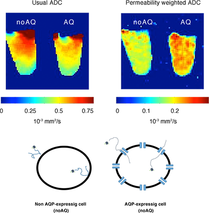
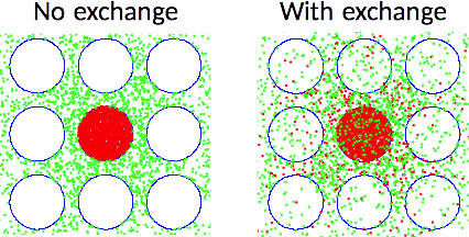
Figure 1-2 Monte-Carlo simulation of water diffusion
Computer simulation is performed to assess the hypothesis of the relation between MRI measurements and permeability of the cell membrane. In this simulation model, water molecules (dots) move around at random. When they occasionally run into the cell membrane (circles), they are either reflected or they permeate depending on a certain probability.
2Technical innovation
This section aims to develop new technology for better clinical imaging output. A compact PET-MRI system for easily plugging into clinical MRI has been developed. New concepts of MR elastography have been proposed and applied to human trials. Noise-reduced and stable analysis methods for multi-strength DWI (DTI, multi-component model, and so on) have been proposed. New methods for easier and more accurate analysis in dynamic-contrast-enhanced MRI have also been developed.
Figure 2-1 Development of compact PET-MRI
A compact PET-MRI system that is compatible with commercial MRIs has been developed. The PET-detector is not built into the gantry of the system but is combined with the RF coil (collaboration with Dr. Yamaya's lab at NIRS and Dr. Suga's lab at Chiba Univ.).
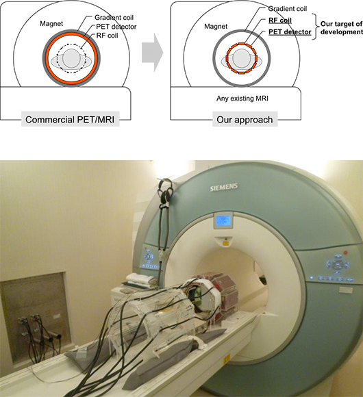
Figure 2-2 Development in MR elastography
Conventional MR elastography (MRE). Wave images can be obtained to synchronize an external oscillator with motion-sensitizing gradient (MSG), which is used to create the elastogram. An oscillator-less MRE has also been developed by our team (collaboration with Dr. Suga's lab at Chiba Univ.).
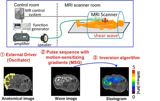
Figure 2-3 Optimization of DWI-parameter calculation for clinics
A new robust method (eDKI) to estimate axial and radial diffusional-Kurtosis imaging maps was developed. The indicated maps are radial K maps obtained by the conventional and developed methods from the same DWI data of 5 different b-values and 21 encoding directions, respectively. The map quality was greatly improved without largely trading off its accuracy. (Microsoft Windows platform software to calculate the eDKI map is available in the "DOWNLOAD APP" section of this website.)
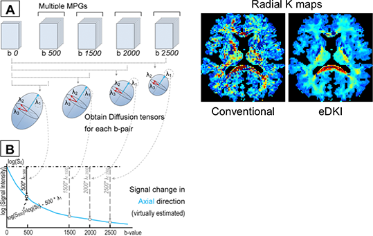
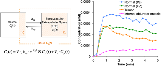
Figure 2-4 Advanced analysis of DCE-MRI
The compartment model (left) is used for pharmacokinetic analysis of dynamic-contrast-enhanced MRI (DCE-MRI). Blood vessels and EES are expressed with one compartment, respectively, and kep represents the rate constant between the compartments. The parameters were calculated by fitting the model to the time-concentration curve (right) (collaboration with Dr. Ikoma's lab at NIRS).
3Clinical application
Clinical application: Applying new evidence-based technologies to clinical imaging is the purpose of this section. Comparative study between US and MR elastography has been performed. Evidence-based biomarkers (DWI, DCE-MRI, MR spectroscopy) have been applied to cancer research with radiation therapy and pediatric medicine. Neuro-scientific MR techniques have been applied to psychiatric fields such as cognitive behavior therapy.
Figure 3-1 Assessment for the optimized application of MRE / USE to clinics
A phantom study was performed to analyze the accuracy of shear-wave-velocity (SWV) measurement of MR and US elastography when compared with a rheometer. (Left) The map indicates the SWV of the phantom that was measured by rheometer. The bottom of the phantom indicates faster SWV (indicating firmer structure) because the area is compressed by the weight of the phantom itself. (Right) The SWV of US elastography (VTQ) and MRE indicate a strong correlation with that of a rheometer.
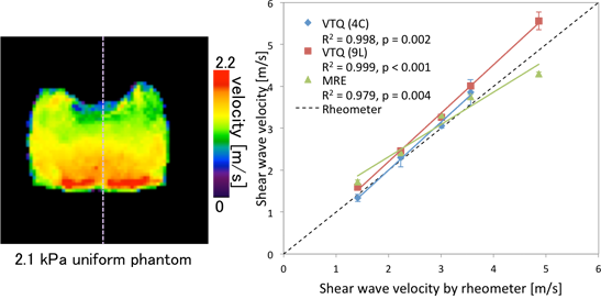
Figure 3-2 Clinical assessment of multiple b-value DWI
DWI parameters were compared in tumors before and after charged particle therapy of cervical cancer. Parameters obtained from non-Gaussian fitting model were more sensitive to the early changes than the usual apparent diffusion coefficient (ADC).
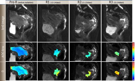
Figure 3-3 Compartment model analysis for DCE-MRI in clinics
The model (see section 2-4 of this page) was used for DCE-MRI of prostate cancer. T2-weighted image of the prostate (left) and kep map obtained by the compartmental analysis of DCE-MRI images (right). Increase in kep was observed in the tumor region indicated by arrows (collaboration with Dr. Ikoma's lab at NIRS).
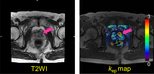
Figure 3-4 MRS spectroscopy and neonatal brain development
The concentrations of metabolites in neonatal brain were measured by MR spectroscopy. The concentration of N-acetylaspartate was well correlated with postconceptional age, which possibly reflected brain development (collaboration with Dr. Aida's group at Kanagawa Children's Medical Center).
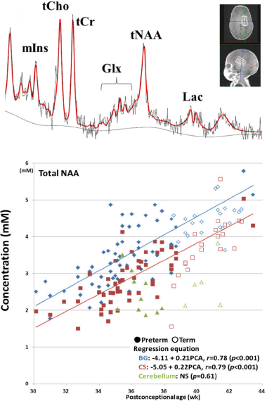
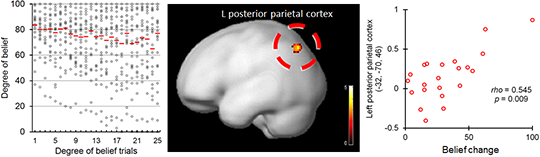
Figure 3-5 Functional brain mapping and neuropathology
Functional brain mapping research for neuropathology of anxiety-related disorders and therapeutic mechanisms of psychotherapies (collaboration with Dr. Shimizu's lab at Chiba Univ.). Belief changes and brain activity correlated with wavering in belief were assessed.